
The musculoskeletal system allows your horse to move around in their environment. The anatomical design of the bones and muscles of the horse reflect the unique needs of the species.
- Intricate system of strong, light-weight tendons in the limbs to allow fast movement of the horse’s large body
- Sophisticated hoof design to absorb the pressures caused by the weight of the horse while moving
- Stay apparatus , a sophisticated network of muscles and tendons of the front and hind limbs, allows the horse to stand for long periods of time with minimal effort. The ability to rest while standing allows the horse to escape faster if a predator arrives unexpected.
Abnormalities of the musculoskeletal system include injuries (acute or chronic) and/or conformation problems.
- Conformation abnormalities
o Congenital: Angular limb deformities, Clubfoot
o Acquired: Knock knees, Bow legged, Cow-hocked; Upward fixation of the patella
- Degenerative conditions
o Osteochondritis dissecans (OCD)/Osteochondrosis: Shoulder; Stifle; Hock
o Osteoarthritis develops in overly stressed joints over time. Bucked shins. Splints. Bone Spavin.
o Patellar chondromalacia
- Fractures
o Forelimb: Humerus; Radius/Ulna; Carpus; Metacarpals; Splint bones; Sesamoid; First phalanx; Second Phalanx; Third Phalanx; Navicular bone
o Hindlimb: Femur; Patella; Tibia; Tarsus, Calcaneus; Metatarsals; Splint bones; Sesamoid; First phalanx; Second Phalanx; Third Phalanx; Navicular bone
o Spine: Cervical; Thoracolumbar; Sacrum; Coccyx
- Hoof problems
o Corns, Cracks, Quittor, Sheared heels, Sidebone
o Laminitis (Founder)
o Sole abscess, bruise
- Infectious diseases : viral, bacterial, Rickettsial; septic arthritis; osteomyelitis
- Metabolic and nutritional disorders predispose to injury
- Soft tissue (muscle and/or tendon) injuries and inflammatory conditions
o Forelimb: Bicipital bursitis, Sweeney, Superior check ligament strain, Bucked shins, Suspensory Ligament Desmitis/Rupture, Inferior Check Ligament Desmitis, Bowed tendons, Sesamoiditis, Chronic proliferative synovitis, Osselets, Ringbone, Pedal osteitis, Buttress foot, Navicular syndrome/disease
o Hindlimb: Hip dislocation; Round ligament rupture; Whorlbone lameness (Trochanteric bursitis); Myopathy: fibrotic and ossifying; Meniscal Tears (stifle); Peroneus Tertius Rupture; Thoroughpin (DDF tenosynovitis), Curb (Tarsal Plantar Desmitis); Cunean Bursitis; Bog Spavin; Stringhalt; Capped hock; Hock dislocation
o Spine: Sacroiliac dislocation
o Myositis: bacterial, viral
o Exertional Rhabdomyolysis (Monday Morning Sickness)
- Cancer
Surgical intervention may be needed in some cases of musculoskeletal disease. Recovery from musculoskeletal disease or injury requires a combination of pain management, farrier work, exercise restriction, rehabilitatitive care, and nutritional evaluation.
Carpus

The carpal joint (the “knee) rests between the radius and cannon (third metacarpal) bone. Lameness associated with the carpal joint can include problems within the joint, the surrounding bones, and the tendons and ligaments that pass over. Symptoms of carpal joint disease vary from mild lameness to declining athletic performance to severe lameness. Physical examination findings may show swelling and heat surrounding the carpal joint spaces, pain on manipulation, and decreased range of motion. The strong carpal joint withstands tremendous amounts of pressure and, over time (particularly in race horses) these repetitive forces can lead to osteoarthritis. Other carpal joint abnormalities include: osteochondral fragments, fractures to the carpal bones, and soft tissue inflammation (e.g.: intercarpal ligament tears, suspensory desmitis).
Coffin Joint
The coffin joint lies between the second and third phalanges of each limb. The joint space is located under the interface of the hoof and haired skin on the limb (coronet band). Horses with coffin joint pain present with a wide range of symptoms: from acute lameness to poor athletic performance. The forelimbs are more commonly affected. Physical examination findings include coffin joint distension and pain on joint flexion. The most common causes of lameness in the coffin joint are synovitis (inflammation of the joint capsule) and osteoarthritis. Other abnormalities include: trauma, ligament inflammation (desmitis), and fractures to the phalanx bones.
Distal Limb

The musculoskeletal system allows your horse to move around in their environment. The anatomical design of the bones and muscles of the horse reflect the unique needs of the species.
- Intricate system of strong, light-weight tendons in the limbs to allow fast movement of the horse’s large body
- Sophisticated hoof design to absorb the pressures caused by the weight of the horse while moving
- Stay apparatus , a sophisticated network of muscles and tendons of the front and hind limbs, allows the horse to stand for long periods of time with minimal effort. The ability to rest while standing allows the horse to escape faster if a predator arrives unexpected.
Elbow Joint

Horses bear 60% of their body weight in their front limbs, thus elbow health is essential. For normal elbow function, all of the bones (humerus, radius, and ulna) of the joint must align. Problems in the horse’s elbow often stem from trauma: humerus and olecranon (tip of the elbow) fractures after being kicked or falling. Horses with elbow pain often do not bear weight on the affected limb; the elbow may appear dropped in position. A healthy body weight is the best insurance to minimize joint inflammation in your horse. Anti-inflammatory medications, nutritional supplements, physical therapy, and/or surgery may be recommended by your veterinarian to keep your horse’s elbow joints strong and mobile.
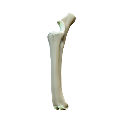
Femur

The large femur bone connects the hip and stifle joints. Large muscle groups attach to the femur to allow flexion and extension of the rear limb. Abnormalities associated with the femur commonly occur from trauma. Trauma to the femur most commonly results in a fracture; dislocation of the femur from the hip joint occurs infrequently. Horses with femur fractures are visibly lame and cannot bear weight on the limb. Diagnosis of a femur fracture is based on x-ray findings. Fractures in foals may be able to be corrected surgically. Unfortunately, femur fractures in adults carry a poor prognosis for return to function and euthanasia is often recommended.
Fetlock Joint

The fetlock joint, or ankle, rests between the cannon (third metacarpal) and the long pastern (first phalanx) bones. Tucked behind the fetlock joints are a pair of sesamoid bones encased in the flexor tendons. Horses with fetlock joint pain present with a wide range of symptoms: from acute lameness to poor athletic performance. The forelimbs are more commonly affected. Physical examination findings include fetlock joint distension, heat, and pain on joint manipulation. The most common causes of lameness in the fetlock joint are synovitis (inflammation of the joint capsule), suspensory ligament injury, luxations, osteochondrosis, and fractures (especially in athletic horses). Other abnormalities include: osteochondrosis, trauma, and ligament injury/inflammation.
Hip Joint

The hip joint connects the hind limb to the pelvic girdle. The hip joint is a ball (top of femur) and socket (acetabulum of the pelvis) joint that is surrounded by large muscle groups for stabilization. Detection of pain in this joint is uncommon except in horses with fractures of the acetabulum, often caused by trauma. Even in cases of fracture, the lameness can be subtle. Other causes of hip discomfort, while uncommon, include osteoarthritis, hip dislocation, and injury to the proximal femur.
Hock

div p The hock joint rests between the tibia and the third metatarsal (cannon) bone. The bones of the hock joint line up in 3 rows and interconnect by numerous ligaments to stabilize the joint. Horses with hock joint pain present with a wide range of symptoms from acute lameness to poor athletic performance. Physical examination findings include distension of the hock joint, heat, and pain on joint manipulation. The most common causes of lameness in the hock joint are osteoarthritis (i.e. bone spavin), synovitis (inflammation of the joint capsule, bog spavin), osteochondrosis, desmitis (inflammation of a ligament, curb), and fractures (especially in athletic horses). p div
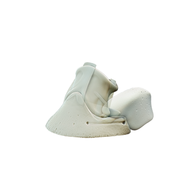
Hoof

The sophisticated equine hoof design absorbs the pressures and distributes the weight of the horse while moving. Trauma to the foot commonly leads to infection, pain, and disuse. Other contributors include poor substrate conditions (excessive moisture or dryness) and nutrition (e.g.: grain overload, sudden dietary changes). Horses with foot problems may or may not show signs of lameness. Feet need to be cleaned out daily to inspect for hoof wear abnormalities and trauma. Any problems detected should be evaluated by your veterinarian and farrier to ensure the best outcomes.
Metacarpus

The metacarpal bone (also known as the cannon bone) is one of the strongest bones of the horse’s body. This bone rests between the carpus (knee joint) and the fetlock joint. Running along the back of this bone are the major flexor tendons of the distal limb. Horses with metacarpal pain present with a wide range of symptoms: from acute lameness to poor athletic performance. Physical examination findings include swellings, heat, and pain on palpation of the bone and/or tendons. The most common causes of lameness in the metacarpal region include: osteochondral fragmentation, bucked shins in athletic horses, fractures, desmitis (inflammation of the suspensory and check ligaments), and tendon injuries.
Pastern Joint

The pastern joint rests between the proximal and distal phalangeal bones. Horses with pastern joint pain present with a wide range of symptoms: from acute lameness to poor athletic performance. The forelimbs are more commonly affected. Physical examination findings include pastern joint distension, heat, and pain on joint manipulation. The most common causes of lameness in the pastern joint are synovitis (inflammation of the joint capsule), luxations, and fractures (especially in athletic horses).
Pelvis

The pelvis is composed of 4 bones (ilium, ischium, pubis, and sacrum) that form a box to connect the spine to both of the hind limbs at the hip joints. The pelvic box surrounds and protects portions of the intestinal, urinary, and reproductive tracts. Fractures from trauma (e.g.: falls, slips, doing “the splits”) represent the most common cause of injury to the pelvis. Due to the box-like nature of the pelvis, fractures tend to be multiple and concomitant injury to the internal organs may occur. Horses with hip pain rise more slowly or limp; the hips may appear uneven. A healthy body weight is the best insurance to minimize joint inflammation in your horse. Anti-inflammatory medications, nutritional supplements, physical therapy, and/or stall confinement may be recommended by your veterinarian to keep your horse’s hip joints strong and mobile.
Radius Ulna
The radius and ulna shape the forearm: they lie distal to the elbow and above the carpus (“knee”). These bones are covered on three sides by muscle groups that allow flexion and extension of the distal forelimb. Injury to the radius and ulna is uncommon, but cause a non-weight bearing lameness characterized by a dropped elbow. Physical examination findings include swellings, heat, and pain on palpation of the bone and/or muscle groups. Fractures of the radius and ulna occur infrequently in the horse.
Shoulder Joint

Horses bear 60% of their body weight in their front limbs; thus, shoulder health is essential. The shoulder connects the front limbs to the trunk and provides support for the front half of the body. For normal function, all of the bones (humerus and scapula) of the shoulder joint must align properly. Disruption of the normal gliding of the shoulder joint results in pain. Developmental abnormalities (e.g.: osteochondritis dissecans, OCD) and trauma to the shoulder (e.g.: fractures) are common causes of pain in the shoulder. Without intervention, arthritis progresses in the joint. Shoulder pain may result in poor performance; some horses may limp on the affected leg. A healthy body weight is the best insurance to minimize joint inflammation in your horse’s shoulders. Anti-inflammatory medications, nutritional supplements, physical therapy and/or surgery may be recommended by your veterinarian to keep your horse’s shoulder joints strong and mobile.
Skull

The skull, containing numerous fused bones, gives shape to your horse’s head, facilitates jaw movement, and envelops the fragile brain to offer protection from trauma. Skull injuries commonly occur from head trauma resulting in fractures, brain injury, and bleeding. Built within the skull are bone encapsulated air pockets known as sinuses. The sinuses moisten inhaled air and trap inhaled particles within secreted mucous. Horses with sinus discomfort develop a nasal discharge. Sinus problems occur with bacterial and viral infections (e.g.: Equine Herpesvirus), allergies, and tooth problems.
Stifle

Your horse loves to run…healthy joints help ensure that this activity can continue. The stifle joint is a common site of injury in horses. Stifle injuries may involve bony (e.g.: osteochondrosis dissecans) or soft tissue (e.g.: ligament sprains/ruptures). Horses with stifle pain may hold up the affected leg or partially bear weight on it. A healthy body weight is the best insurance to minimize joint inflammation in your horse. Anti-inflammatory medications, nutritional supplements, physical therapy, and/or surgery may be recommended to keep the stifle joints strong and mobile.
Tibia Meniscus

The tibia rests between the stifle and hock joints. This long bone is surrounded on three sides by muscle groups that allow flexion and extension of the distal limb. Horses with tibial injuries present with a wide range of symptoms: from acute, non-weight bearing lameness to poor athletic performance. The most common causes of tibial injury include fractures, meniscal tears, rupture of the peroneus tertius, and tenosynovitis of the deep flexor tendon sheath.
What's Next
Call us to schedule an appointment
Meet with a doctor for an initial exam.
Put a plan together for your pet.
